This information is of a general nature only. Please consult your doctor for specific advice concerning your condition.
What is Stereotactic Radiosurgery?
Stereotactic radiosurgery is an alternative to surgery and is used to treat some brain tumours. It involves the focusing of multiple pinpoint radiation beams very precisely onto a brain tumour to neutralise or destroy the tumour. Because these beams come from many directions and only intersect at the tumour there is very little dose received by surrounding brain.
The treatment is painless (you can’t hear or feel the radiation) and in many instances is delivered on a single occasion. In some instances this form of precise radiation is delivered in multiple doses over a number of days in which case we call it “fractionated stereotactic radiotherapy”.
No incision is made in the head as the radiation beams come from external to the skull.
A/Prof Jonker is the only neurosurgeon in Australia to have completed an overseas fellowship specifically dedicated to stereotactic radiosurgery (UCLA, Los Angeles 2009) and has performed well over a thousand of these procedures including both LINAC systems and Gamma Knife. He is considered an expert and has been an invited speaker on this topic at multiple national and international meetings.
In addition to brain tumours this technique can be used to treat vascular malformations of the brain, trigeminal neuralgia and some cases of tremor or Parkinson’s Disease.
If the treatment uses radiation and not a scalpel, why is it called radiosurgery?
There are 3 reasons why it is called radiosurgery –
Firstly, there is an emphasis on exactly targeting a tumour and avoiding injury to any surrounding structures, similar to traditional open surgery.
Secondly, radiosurgery is usually a single dose, one-off treatment – again, similar to surgery.
The third reason is historical. Radiosurgery was devised by a neurosurgeon, who conceived of a method of doing neurosurgery where the X-ray replaced the scalpel as a form of knife.
What conditions are suitable for radiosurgery treatment?
The most commonly treated tumours are secondary brain tumours (also known as metastases or mets), acoustic neuromas, meningiomas, and pituitary adenomas. There are a couple of non-tumour conditions which are also commonly treated called arteriovenous malformations (AVMs) and trigeminal neuralgia.
Other less common reasons for treatment include gliomas, craniopharyngioma, glomus tumours, cavernous malformations, chordomas and chondrosarcomas, tremor and epilepsy.
Radiosurgery is not suitable for all patients, so it is best to visit a team that has both neurosurgery and radiation oncology expertise.
What are the common names of radiosurgery systems?
Names such as Cyberknife, Gamma Knife and Zap-X refer to systems specifically developed for radiosurgery use, but other units use Varian or other equipment.

Zap-X Gyroscopic Stereotactic Radiosurgery
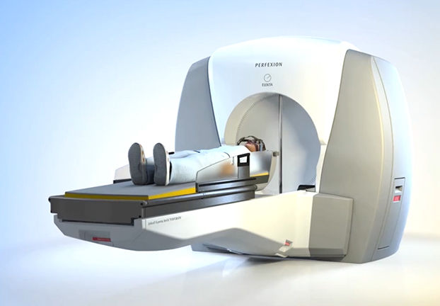
Gamma Knife
Do patients require an anaesthetic or admission to hospital?
No, patients receive their treatment whilst awake – and can return home the same day.
What can I expect after the treatment?
It is common to feel a mild headache or some fatigue for a few days afterward. Usually, no special medications are required. Many patients will return to work the day following treatment. Most patients don’t lose any hair with this treatment, but occasionally patients will lose a very small amount of hair.
Framed versus frameless?
Traditionally radiosurgery has required the application of a head frame to achieve the precise positioning of the patient (and their tumour) for their treatment.Some of the modern radiosurgery devices have methods of achieving accurate positioning of the patient using image guidance systems on the radiosurgery machine. A mask is used in these patients.
What complications can occur?
This is always best discussed in the context of the patient’s individual condition.
Is radiosurgery for patients that are not good candidates for surgery?
Patients who are not good candidates for surgery may still be good candidates for radiosurgery. However, having radiosurgery does NOT mean that surgery was not possible – in many cases it simply means that the radiosurgery was either safer or more effective than surgery. Simply put, sometimes radiosurgery is just a better treatment for a condition than surgery.
The technical side
In radiosurgery the aim is to deliver the dose precisely and sharply to the tumour with minimal spreading of the dose into the adjacent structures which might include the brain or important nerves. Several physical factors are known to affect how well this can be done.
Cobalt versus linear accelerator (LINAC). There are 2 methods used for creating the high energy photons needed for radiation treatment. The first is radioactive decay of Cobalt 60 (seen in the Gamma Knife). The second is a device called a linear accelerator (used by other systems). Cobalt 60 always produces photons of 2 specific wavelengths. Linear accelerators produce a spectrum of photons at various energies. Whilst there is no specific benefit between cobalt and LINAC for radiation production per se the energy levels of the photons in Gamma Knife and the new Zap-X linear accelerator are of a similar energy level to each other and are known to produce a sharper edge to the radiation dose than the Cyberknife or other linear accelerators.
Source to Axis Distance (SAD) – the distance between the radiation source and the target affects the sharpness of the radiation dose, with closer distances being superior. Conventional linear accelerators used for radiosurgery have an SAD of 100cm, but the Gamma Knife and Zap-X have an SAD of 40-45cm.
Collimation – this is the process of narrowing and aligning the radiation beams so that they can be focused on the target. Collimation can be subject to leakage which can reduce the sharpness of the beam and allow more dose to the body or healthy brain. For a standard linear accelerator this can be around 1.3% whereas the Zap-X and Gamma Knife are 0.001%. The smallest collimator size (for the finest work) is 4mm for the Gamma Knife and 3mm for the Zap-X.
Shielding – Typically radiation machines are housed in a bunker to prevent radiation spreading out of the room. The Zap-X is different to all other devices in that the machine itself has its own radiation shield so there is no need for other people such as the treating team to leave the room during treatment.
Fixation – A stereotactic head frame has been the historical gold standard for fixing the patient’s head in 3 dimensional space during treatment. Other systems use a mask and some form of X-rays to position the head. Whilst the latter system has been shown to rival the accuracy of the head frame when the patient first goes into machine, the problem can be slight drifts of the head within the mask during treatment, as time goes on. The Zap-X overcomes this by being the only radiosurgery device that makes slight adjustments by a tiny fraction of a millimetre every 45 seconds during treatment to make sure there is no significant drift of the patient.


