Vestibular Schwannomas, also known as Acoustic Neuromas, are benign tumours that arise from balance nerves at the base of the brain.
Symptoms
The balance nerves travel with the hearing nerve and the facial nerve so common symptoms are vertigo (dizziness), hearing loss (on one side), tinnitus (ringing in the ear). Facial weakness doesn’t usually occur from the tumour but is a major focus in the treatment decisions.
Deciding on a treatment strategy
When a patient is faced with decisions regarding treatment of an acoustic neuroma (vestibular schwannoma) making the right choice is not always easy and straightforward. This is in part because treatment options are very different from each other and include –
- Surgery
- Stereotactic radiosurgery or fractionated stereotactic radiation treatment
- ‘Wait and see’
Another reason that the decision in not simple when looking at the literature has to do with the very long follow-up periods that are necessary to establish the behaviour of what is usually a slow growing benign tumour and the effects of treatment. Finally, there are relatively few people (at least in Australia) who have experience in all of the treatment types, so invariably the advice you get may be skewed toward one approach rather than another- and you may receive conflicting opinions from different sources.
History
The first successful resection of an acoustic neuroma was in the 1890’s – often attributed to Charles Ballance (1894). When acoustic neuroma surgery was first performed it was literally life saving. Thoughts of normal facial power and hearing preservation simply didn’t come into the equation. Likewise the instrumentation was fairly primitive- the surgeons finger was used to sweep the tumour out!
Over time surgical techniques have allowed for more frequent preservation of the facial nerve. This depends on size of the tumour and surgical experience. Brackmann (from the very experienced House Clinic) has reported a 17% chance of significant facial paralysis (House Brackmann Grade 3-6) amongst patients with a 1.5cm-2.5cm tumour operated on using a translabyrinthine approach versus a a risk of 9% of significant facial paralysis if the tumour is less than 1.5cm (1). For the 1.5-2.5cm facial power was completely normal in 71% of the patients. These results were after 1 year of follow-up so the results can be considered to be the stable ‘going forward’ rates.
Pollock and co-authors from the Mayo Clinic are, to the best of my knowledge, the only group that have used an independent observer (who didn’t know what type of treatment the patient had) to assess the facial nerve function and found it to be normal at one year following surgery in 69% of patients (2). Their group included patients with small and midsize tumours deemed to be suitable for either radiosurgery or surgery. These tumours averaged 14mm in diameter.
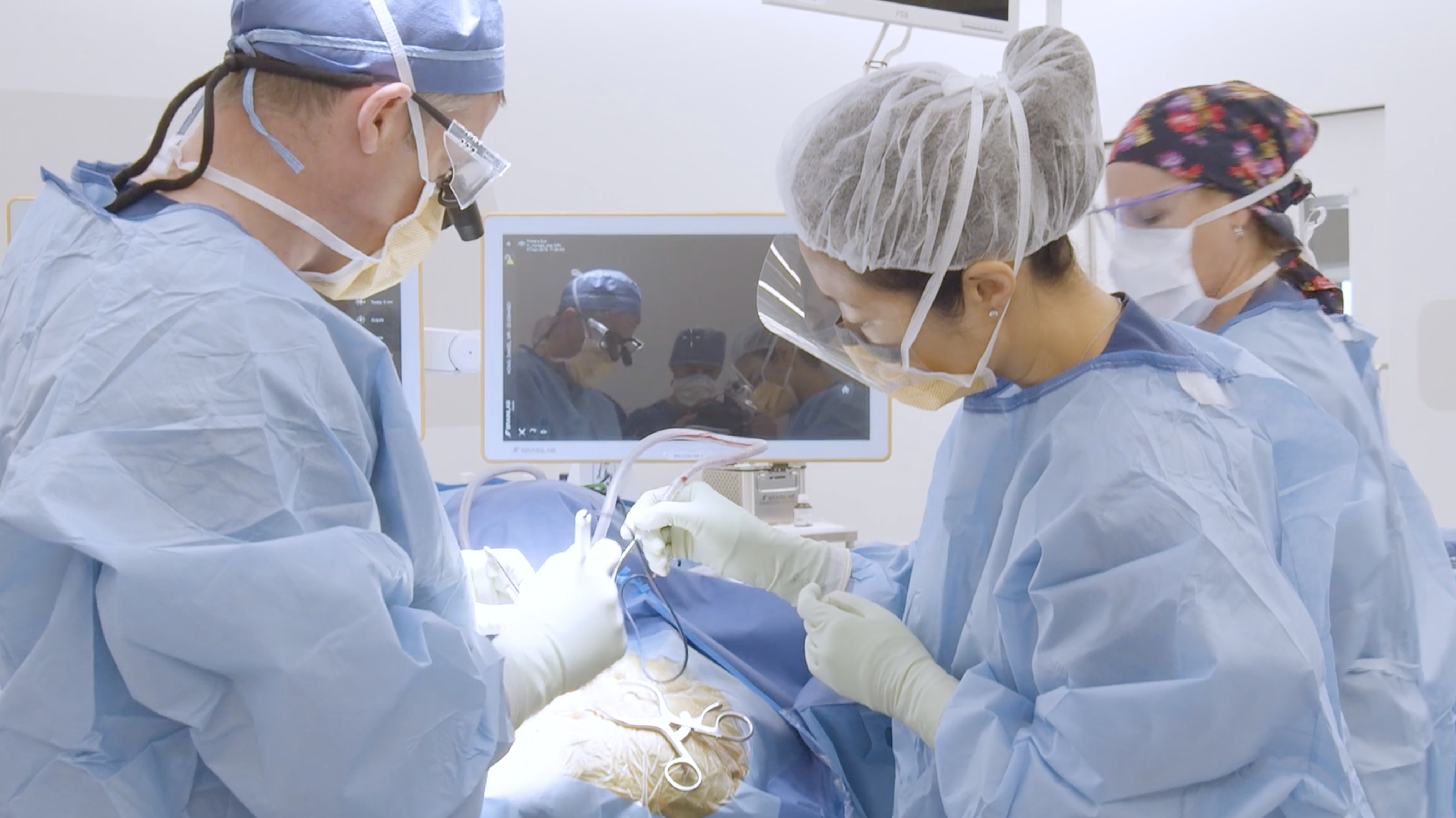
How is surgery performed?
Surgery for vestibular schwannoma is always performed under a general anaesthetic (patient asleep). The 2 approaches commonly used by Dr Jonker (in association with his ENT surgery colleagues) are the retrosigmoid and the translabyrinthine approach. The retrosigmoid approach involves less bony drilling and in some cases hearing can be preserved (more about this below). The translabyrinthine involves a more extensive amount of bony drilling but provides a more direct approach to the tumour. The best approach for an individual patient will be dictated by the anatomy and their current hearing. Occasionally, in certain circumstances, a cochlear implant or brainstem implant will be inserted at the time of surgery for hearing rehabilitation. These surgeries are 2 surgeon operations with the collaboration of the neurosurgeon and the ENT surgeon. Much of the surgery is focussed on protecting the facial nerve which is often thinned out by long term compression from the tumour.
What about preservation of hearing with surgery?
This is a little more difficult to assess since some approaches to surgery (ie translabyrinthine) will definitely cause deafness on that side. Samii from Hannover in Germany has reported his experience with hearing preservation using the retrosigmoid approach. He managed to achieve a very impressive 51% preservation of functional hearing in his more recent cases.
What is radiosurgery?
Stereotactic radiosurgery is a technique pioneered by a neurosurgeon (and later refined and developed by other neurosurgeons) which aims to give a high dose of radiation, usually as a single treatment (ie all done on one day) shaped to fit the target, with a very rapid dose fall-off at the periphery so as to avoid significant dose to any surrounding structures.
For a patient with an acoustic neuroma this means delivering a single dose of radiation the same 3-D shape as your acoustic neuroma- accurately to the tumour, while avoiding significant dosage to the brainstem, sensory nerves to the face, cochlea and so forth.
Radiosurgery is not new. Patients have been treated with this technique since 1967. The technology has been continually refined, however. In the 1990’s there was a shift towards using a lower dose to treat acoustic neuromas so most of the relevant medical literature comes from the last 10-20 years for this particular tumour type.
The aim of the treatment is tumour control. This means that the tumour may or may not shrink or change its appearance (for example less contrast enhancement on the MRI) – but as long as it doesn’t increase in size it is considered to be controlled.
How is the precise 3-D dose created? There are different techniques for doing this. Firstly the radiation need to either be emitted or created- this is either done using radioactive decaying cobalt or using a device called a linear accelerator. Some radiosurgery devices use linear accelerators, and one uses cobalt. It doesn’t matter which technique is used – the net effect is the same. This radiation is then directed toward the tumour from many different angles, and each ‘beam’ overlaps with the others at the site of the tumour. That way there is a high dose on the tumour but only a relatively low dose elsewhere.
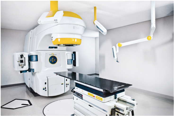
Novalis TX at Chris O’Brien Lifehouse, New South Wales
It would be meaningless to create a precisely shaped dose if it couldn’t be delivered accurately to the correct part of the head- so of course that is an important part of the technology of radiosurgery. In order to do this the tradition has been to use a cranial frame (with pins that screw into the skull to keep it securely fastened). In the past 10 years or so ‘frameless’ radiosurgery has become more commonplace. This uses a mask to immobilize the patient and uses X-rays taken from different angles during treatment to repeatedly check and re-align the patient. Both techniques have similar accuracy- that is about a millimetre or less. We exclusively use the frameless technique at Sydney Radiosurgery (we use a threshold of half a millimetre in any plane to be acceptable, otherwise we realign the patient).
There are various different devices for delivering radiosurgery available on the market- there are some pros and cons of each, and the common names are Novalis, Gamma Knife, and CyberKnife. These 3 have been compared in their ability to create a 3 dimensional dose conforming to the target, and provide a rapid dose fall-off at the periphery and have been found to be the same as each other (3,4).
What is fractionated stereotactic radiotherapy?
Conventional radiotherapy has been less precise in its ability to focus the treatment on a tumour but has achieved success by spreading the dose out over multiple days (called fractionation). This relies on the tumour being less capable of repairing itself after each treatment than the nearby normal tissue is (which has more capability of ‘bouncing back’ from each dose).
Fractionated stereotactic radiotherapy combines the benefits of the precise 3 dimensional dose being applied to the tumour, with the additional advantage of sparing normal tissue even further by spreading the treatments out over time.
Why would this be an advantage if radiosurgery is already so accurate that nearby structures can be avoided?
Acoustic neuroma is a very particular type of tumour. By definition the facial nerve (for facial muscle power) and cochlear nerve (for hearing) are draped over the surface of the tumour. No matter how accurately the radiation is given, the dose prescribed to the surface of the tumour will be given to that portion of those nerves.
It turns out that the facial nerve is relatively robust to radiation, but the cochlear (hearing) nerve is what is known as a special sensory cranial nerve and (like the nerves for vision) is more susceptible to radiation. The main reason for considering fractionated stereotactic radiotherapy is that there are a number of reports in the literature that indicate that whilst the overall tumour control rate and facial nerve injury rate are the same between fractionated stereotactic radiotherapy and stereotactic radiosurgery, the hearing preservation rate appears slightly higher with fractionated stereotactic radiotherapy (5,6,7).
Fractionated stereotactic radiotherapy is typically given over 28 days – with weekends off- so it takes just under 6 weeks.
Stereotactic radiosurgery and fractionated stereotactic radiotherapy results
In 2009 I summarized the results in the literature as part of a project presented as a poster at the World Society of Stereotactic and Functional Neurosurgery meeting in Toronto. Since these results represent a summary of large numbers of patients I will present them here.
The risk of significant facial paralysis with stereotactic radiosurgery was found to be 0.4% (range 0-2%) in 7 studies of 2625 patients undergoing stereotactic radiosurgery for acoustic neuroma (2,8-13).
The chance of hearing preservation was found to be 55% (range 38-80%) in 3 studies involving 364 patients undergoing stereotactic radiosurgery for acoustic neuroma (8,14,15).
The chance of obtaining tumour control with radiosurgery was 97.7% (range 94.2%-98.6%) in 7 studies of 2089 patients undergoing radiosurgery (8,10,14,16-19).
The results for fractionated stereotactic radiotherapy appear to be similar for control of tumour and facial nerve preservation- however hearing preservation is improved. This has been documented by a number of centres with hearing preservation rates of 68-94% (7)(5,20).
How we approach vestibular schwannoma
For a new diagnosis of tumour, or no documented enlargement on serial imaging we generally recommend the ‘watch and wait’ approach since there is good evidence that many of these tumours do not enlarge over significant time intervals (21).
If the tumour is moderately large (ie 2.5-3 cm) treatment would be considered, and surgery generally preferred.
If the tumour has caused some unilateral (one sided) hearing loss but there is still functional hearing (hearing worth saving) in the affected ear, we would consider offering treatment with fractionated stereotactic radiotherapy on the basis that the hearing preservation rates may be better with this treatment than with watching alone. This is always a detailed discussion, since the evidence is sparse and we cannot be dogmatic about this.
For patients with documented enlargement of a tumour less than 25mm we would generally recommend stereotactic radiosurgery if they have functional hearing, and single fraction treatment if no functional hearing or the hearing is less important to the patient.
Occasionally patients with tumours >3cm are otherwise unsuitable for surgery but need treatment- in this case we consider fractionated stereotactic radiotherapy.
As always there are many more considerations to take into account, not least the preference of the patient! As always the treatment needs to be tailored to your individual situation and the discussion here is general.
References
- Brackmann DE, Cullen RD, Fisher LM. Facial nerve function after translabyrinthine vestibular schwannoma surgery. Otolaryngology–head and neck surgery : official journal of American Academy of Otolaryngology-Head and Neck Surgery. 2007 May 1;136(5):773-777.
- Pollock BE, Driscoll CL, Foote RL, Link MJ, Gorman DA, Bauch CD, et al. Patient Outcomes after Vestibular Schwannoma Management: A Prospective Comparison of Microsurgical Resection and Stereotactic Radiosurgery. Neurosurgery. 2006 Jul. 2;59(1):77-85.
- Ma L, Sahgal A, Descovich M, Cho Y, Chuang C, Huang K, et al. Equivalence in dose fall-off for isocentric and nonisocentric intracranial treatment modalities and its impact on dose fractionation schemes. Int J Radiat Oncol Biol Phys. 2010 Mar. 1;76(3):943-948.
- Schoonbeek A, Monshouwer R, Hanssens P, Raaijmakers E, Nowak P, Marijnissen JPA, et al. Intracranial radiosurgery in the Netherlands. A planning comparison of available systems with regard to physical aspects and workload. Technol Cancer Res Treat. 2010 Jun. 1;9(3):279-290.
- Andrews DW, Werner-Wasik M, Den RB, Paek SH, Downes-Phillips B, Willcox TO, et al. Toward dose optimization for fractionated stereotactic radiotherapy for acoustic neuromas: comparison of two dose cohorts. Int J Radiat Oncol Biol Phys. 2009 Jun. 1;74(2):419-426.
- Andrews DW, Suarez O, Goldman HW, Downes MB, Bednarz G, Corn BW, et al. Stereotactic radiosurgery and fractionated stereotactic radiotherapy for the treatment of acoustic schwannomas: comparative observations of 125 patients treated at one institution. Int J Radiat Oncol Biol Phys. 2001 Aug. 1;50(5):1265-1278.
- Selch MTS, Pedroso A, Lee SP, Solberg TD, Agazaryan NAE, Cabatan-Awang C, et al. Stereotactic radiotherapy for the treatment of acoustic neuromas. J Neurosurg. 2004 Nov. 1;101 Suppl 3:362-372.
- Chopra R, Kondziolka D, Niranjan A, Lunsford LD, Flickinger JC. Long-term follow-up of acoustic schwannoma radiosurgery with marginal tumor doses of 12 to 13 Gy. Int J Radiat Oncol Biol Phys. 2007 Jul. 1;68(3):845-851.
- Hasegawa T, Fujitani S, Katsumata S, Kida Y, Yoshimoto M, Koike J. Stereotactic radiosurgery for vestibular schwannomas: analysis of 317 patients followed more than 5 years. Neurosurgery. 2005 Aug. 1;57(2):257-65; discussion 257-65.
- Hempel JM, Hempel E, Wowra B, Schichor C, Muacevic A, Riederer A. Functional outcome after gamma knife treatment in vestibular schwannoma. Eur Arch Otorhinolaryngol. 2006 Aug. 3;263(8):714-718.
- Myrseth E, Møller P, Pedersen P, Vassbotn FS, Wentzel-Larsen T, Lund-Johansen M. Vestibular schwannomas: clinical results and quality of life after microsurgery or gamma knife radiosurgery. Neurosurgery. 2005 May 1;56(5):927-35; discussion 927-35.
- Regis J, Roche PH, Delsanti C, Thomassin JM, Ouaknine M, Gabert K, et al. Modern management of vestibular schwannomas. Progress in neurological surgery. 2007;20:129-141.
- Vachhrajani S, Fawaz C, Mathieu D, Menard C, Cusimano M, Gentili F, et al. Complications of Gamma Knife surgery: an early report from 2 Canadian centers. J Neurosurg. 2008 Dec. 1;109 Suppl:2-7.
- Hasegawa T, Fujitani S, Katsumata S, Kida Y, Yoshimoto M, Koike J. Stereotactic Radiosurgery for Vestibular Schwannomas: Analysis of 317 Patients Followed More Than 5 Years. Neurosurgery. 2005 Aug. 2;57(2):257-265.
- Tamura M, Carron R, Yomo S, Arkha Y, Muraciolle X, Porcheron D, et al. Hearing preservation after gamma knife radiosurgery for vestibular schwannomas presenting with high-level hearing. Neurosurgery. 2009 Feb. 1;64(2):289-96; discussion 296.
- Friedman WA, Bradshaw P, Myers A, Bova F, Bradshaw P, Myers A, et al. Linear accelerator radiosurgery for vestibular schwannomas.[see comment]. J Neurosurg. 2006 Nov. 1;105(5):657-661.
- Pollock BE. Management of Vestibular Schwannomas that Enlarge after Stereotactic Radiosurgery: Treatment Recommendations Based on a 15 Year Experience. Neurosurgery. 2006 Feb. 2;58(2):241-248.
- Roche P, Khalil M, Thomassin J, Delsanti C, Regis J. Surgical removal of vestibular schwannoma after failed gamma knife radiosurgery. Progress in neurological surgery. 2008;21:152-157.
- van Eck A, Horstmann G, van Eck A, Horstmann G. Increased preservation of functional hearing after gamma knife surgery for vestibular schwannoma. J Neurosurg. 2005;102 Suppl:204-206.
- Koh E, Millar B, Ménard C, Michaels H, Heydarian M, Ladak S, et al. Fractionated stereotactic radiotherapy for acoustic neuroma: single-institution experience at The Princess Margaret Hospital. Cancer. 2007 Mar. 15;109(6):1203-1210.
- Bakkouri WE, Kania RE, Guichard J, Lot G, Herman P, Huy PTB. Conservative management of 386 cases of unilateral vestibular schwannoma: tumor growth and consequences for treatment. J Neurosurg. 2009 Apr. 2;110(4):662-669.


